Skip to content

Yersinia pestis | Week 2 – Section 2
Transmission
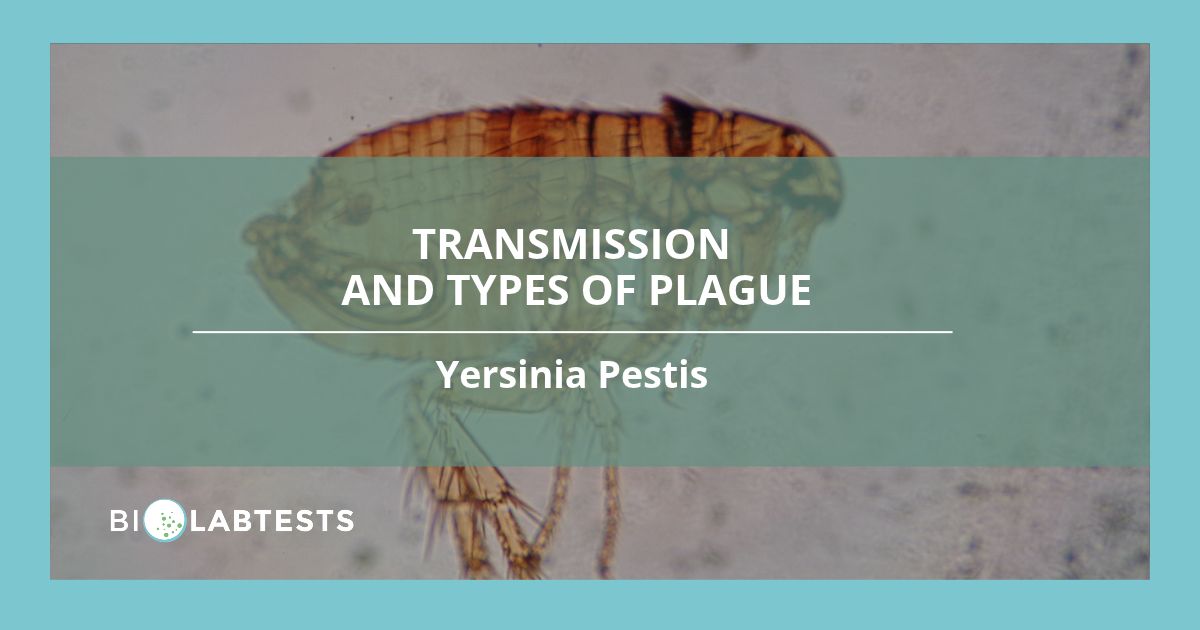
Yersinia pestis is a Zoonotic pathogen – a disease that can transmit from animals to humans or vice-versa – and can be transmitted to humans by being bitten by an infected animal, direct contact with infected animal tissues or through inhalation of aerosolised droplets from infected animals or humans. Animals can also be infected with Y. pestis through contaminated soil (Barbieri et al., 2020). Plague cycles at low rates through animal reservoirs, being transmitted by fleas and mainly infecting wild rodents which can carry the bacteria for longer periods, enabling them to transport the bacteria over longer distances before death but more susceptible hosts can also become infected, amplifying and causing outbreaks of infection (Zeppelini et al., 2016).
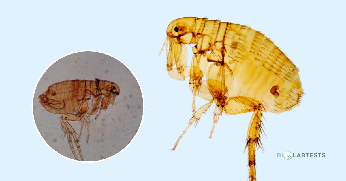
In early phase transmission for a flea, which occurs in the first week after which the flea ingests an infectious blood meal, Yersinia pestis can be transmitted to its next host via the flea biting and taking in subsequent blood meals. However, this mode of transmission has low efficiency and could take numerous infected fleas feeding to achieve the concentration of bacteria needed to become infected (Bosio et el., 2020). The second phase of transmission for a flea is dependent on Yersinia pestis forming a biofilm in the flea's proventricular valve, which connects the flea's oesophagus to the midgut. The biofilm blocks the flea's intake of blood from its blood meals, and the blood attempted to be ingested can come into contact with the biofilm therefore becoming infected before being regurgitated at the bite point to potentially infect the host. This blocking increases the frequency at which the flea attempts to feed and consequently bite to prevent starvation thus increasing the chances for Y. pestis transmission (Barbieri et al., 2020 ; Bosio et el., 2020).
It has been theorised that unlike the third pandemic or modern plague which was/is spread via rats/wild rodents and their vectors that the second pandemic had too high of a household transmission and too few archaeological records of rat distribution and rat epizootics to explain and be the sole cause of the rapid and widespread rate of infection. Instead of the black rat (Rattus rattus) and its flea (X. cheopis), a more direct transmission route has been proposed through human ectoparasites such as lice (Dean et al., 2018).
Forms of plague:
There are five principal forms of plague: bubonic plague, septicaemic plague, pneumonic plague, meningeal plague and pharyngeal plague (Barbieri et al., 2020).
Bubonic plague is the most common form of plague and is characterised by inflammation and swelling in the lymph nodes called buboes, patients will also experience flu-like symptoms such as fever, headache, chills and pain in the affected area (Venugopal & Pechous, 2024). If untreated, bubonic plague can progress and disseminate to the blood (secondary Septicaemic plague) or to the lungs (secondary pneumonic plague).
Septicaemic plague can develop from bubonic plague or pneumonic plague (secondary septicaemic plague) or through direct infection to the blood via a cut (primary septicaemic plague). The bacteria spreads through the bloodstream to colonise secondary tissues and vital organs and can cause multiple organ failure, acute respiratory distress syndrome, disseminated vascular coagulation and gangrene of extremities. Septicaemic plague has a high mortality rate even after antibiotics are administered (Glatter & Finkelman, 2021; Olson & Anderson, 2019; Yang, 2017).
Pneumonic plague can be primary, caused by inhaling aerosolised droplets from other people with pneumonic plague or aerosolised droplets inhaled from infected cats. Secondary pneumonic plague can develop from the person having untreated bubonic plague that has disseminated to the lungs. It has been found that contracting primary pneumonic plague from other people with pneumonic plague usually requires long and prolonged contact (Glatter & Finkelman, 2021).
Pharyngeal plague – a rare form of plague that infects the throat causing pharyngitis and/or tonsillitis, likely developed from either another untreated form of plague, from eating undercooked Y. pestis infected meat or inhaling airborne droplets that lodge in the throat (Barbieri et al., 2020).
Meningeal plague is a rare and often fatal manifestation of plague mostly reported as a complication of primary bubonic plague that can cause focal neurological deficits. Symptoms are similar to other types of meningitis and include nuchal rigidity, headache, and fever (Cooley et al., 2023).
References
Barbieri, R., Signoli, M., Chevé, D., Costedoat, C., Tzortzis, S., Aboudharam, G., Raoult, D., & Drancourt, M. (2020). Yersinia pestis: the Natural History of Plague. Clinical microbiology reviews, 34(1), e00044-19. https://doi.org/10.1128/CMR.00044-19
Bosio, C. F., Jarrett, C. O., Scott, D. P., Fintzi, J., & Hinnebusch, B. J. (2020). Comparison of the transmission efficiency and plague progression dynamics associated with two mechanisms by which fleas transmit Yersinia pestis. PLoS pathogens, 16(12), e1009092. https://doi.org/10.1371/journal.ppat.1009092
Cooley, K. M., Fleck-Derderian, S., McCormick, D. W., & Nelson, C. A. (2023). Plague Meningitis: A Systematic Review of Clinical Course, Antimicrobial Treatment, and Outcomes. Health security, 21(1), 22–33. https://doi.org/10.1089/hs.2022.0081
Dean, K. R., Krauer, F., Walløe, L., Lingjærde, O. C., Bramanti, B., Stenseth, N. C., & Schmid, B. V. (2018). Human ectoparasites and the spread of plague in Europe during the Second Pandemic. Proceedings of the National Academy of Sciences of the United States of America, 115(6), 1304–1309. https://doi.org/10.1073/pnas.1715640115
Glatter, K. A., & Finkelman, P. (2021). History of the Plague: An Ancient Pandemic for the Age of COVID-19. The American journal of medicine, 134(2), 176–181. https://doi.org/10.1016/j.amjmed.2020.08.019
Olson, R. M., & Anderson, D. M. (2019). Shift from primary pneumonic to secondary septicemic plague by decreasing the volume of intranasal challenge with Yersinia pestis in the murine model. PloS one, 14(5), e0217440. https://doi.org/10.1371/journal.pone.0217440
Venugopal, G., & Pechous, R. D. (2024). Yersinia pestis and pneumonic plague: Insight into how a lethal pathogen interfaces with innate immune populations in the lung to cause severe disease. Cellular immunology, 403-404, 104856. https://doi.org/10.1016/j.cellimm.2024.104856
Yang R. (2017). Plague: Recognition, Treatment, and Prevention. Journal of clinical microbiology, 56(1), e01519-17. https://doi.org/10.1128/JCM.01519-17
Zeppelini, C. G., de Almeida, A. M., & Cordeiro-Estrela, P. (2016). Zoonoses As Ecological Entities: A Case Review of Plague. PLoS neglected tropical diseases, 10(10), e0004949. https://doi.org/10.1371/journal.pntd.0004949
How BioLabTests can support you
If you would like to explore our microbiology testing portfolio, please do not hesitate to get in touch with a member of our team on +44 (0) 333 240 8308 or simply complete our contact form.
Please note that BioLabTests does not test human, animal or food samples and we do not test for viruses.